Sofwave vs Ultherapy: where the two devices diverge
Written by the Delight Dermatology editorial team · Medically reviewed by Lead Dermatologist, Delight Dermatology Clinic (Korean Board-Certified Dermatologist, AAD International Fellow, ASLMS) · Last reviewedSofwave and Ultherapy are both ultrasound devices, but they differ in beam geometry and in how many tissue planes they can reach. Sofwave uses seven parallel, non-focused beams to heat a continuous band at one fixed depth of about 1.5 mm in the mid-dermis. Ultherapy uses microfocused ultrasound with visualization (MFU-V) to place discrete thermal coagulation points at selectable focal depths of 1.5, 3.0 and 4.5 mm, with the deepest setting able to reach the SMAS, and it images the tissue in real time before each line is delivered. Both share the 1.5 mm plane. No published head-to-head randomised trial compares the two devices, so any claim that one is simply better than the other is a marketing claim rather than an evidence claim.
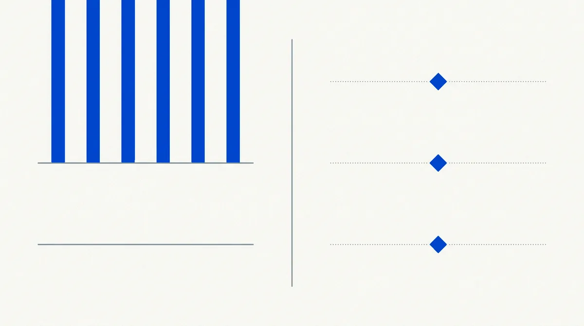
The difference in one picture
Almost everything else follows from where each device deposits energy. The diagram below is drawn to scale on a 0–5 mm axis and holds every other variable constant.
- Sofwave (SUPERB): seven parallel beams, fixed focal depth ≈1.5 mm, thermal zones ≈0.5–2 mm
- Ultherapy (MFU-V): microfocused points at selectable 1.5, 3.0 and 4.5 mm
Epidermis0–0.2 mmPapillary dermis0.2–1 mmReticular dermis1–3 mmSubcutaneous fat3–4.4 mmSMAS≈4.5 mm
Layer boundaries are conventional teaching depths; facial skin thickness varies by site and by patient. The 1.5 mm plane is shared by both devices, which is why the choice between them is made by examination rather than by depth alone.
Side by side
| Aspect | Sofwave (SUPERB) | Ultherapy (MFU-V) |
|---|---|---|
| Beam geometry | Seven parallel, non-focused beams fired synchronously; a continuous heated band | Microfocused beam concentrated to discrete thermal coagulation points along a line |
| Treatment depth | Fixed focal depth ≈1.5 mm; thermal zones ≈0.5–2 mm | Selectable transducers at 1.5, 3.0 and 4.5 mm |
| Deepest plane reached | Mid-dermis. Does not reach the SMAS | The 4.5 mm transducer can reach the SMAS |
| Imaging | No imaging. Contact cooling plus a real-time skin-temperature monitor | Real-time ultrasound visualization of the tissue plane before delivery |
| Frequency | 10–12 MHz as reported in the peer-reviewed series | Device-dependent; lower frequency than Sofwave, consistent with deeper focus |
| Typical session | About 30–60 minutes depending on areas treated | Varies by transducer combination and area; commonly longer for full face and neck |
| Evidence base | Smaller: mostly single-arm series of 14–36 subjects with follow-up to 6–10 months | Larger and older, with more published follow-up across more indications |
| Head-to-head trial | None published. Comparisons between them are indirect. | |
Why depth is the decisive variable
Skin laxity is not one problem. Textural laxity (crepey skin, loss of firmness, fine lines) is largely a dermal issue, and a device that heats the dermis is addressing it directly. Descent of the deeper support structures is a different problem living in a different plane, and heating the dermis does not fix it. A 2026 review of ultrasound in face and neck rejuvenation frames the two technologies exactly this way: MFU-V targets the deep dermis and SMAS at 1.5 to 4.5 mm, while SUPERB delivers at a fixed 1.5 mm to target the mid-dermis, and both are described as suited to mild-to-moderate laxity.
The practical implication is unglamorous. If the examination finds the problem is dermal, a fixed mid-dermal device is well matched to it. If the examination finds significant deeper descent, a device that cannot reach that plane will under-deliver no matter how many passes are used, and the honest options are a deeper modality or a surgical consultation.
What the imaging difference means
Ultherapy's visualization lets the operator confirm the tissue plane and see structures before each line is delivered, which matters most when energy is being placed deep: near bone, near nerve, in areas of variable fat thickness. Sofwave delivers at a shallow fixed depth and manages surface safety with cooling and temperature monitoring instead. Neither approach is a shortcut: they are answers to different risk profiles created by different depths.
Comfort, honestly
The published Sofwave series report mean pain scores of roughly 3.9 to 6.6 on a 0–10 scale depending on the protocol and the area treated. That is a real range, and the upper end is uncomfortable. Ultherapy is also described as painful by many patients. Because no head-to-head study exists, this site will not tell you which one hurts less; that comparison would need a trial that has not been run. What can be said is that comfort is protocol-dependent, that topical anaesthetic is standard, and that pain tolerance is worth raising at the consultation rather than discovering mid-session.
Can they be combined?
They occupy overlapping planes at 1.5 mm, so combination is not a matter of stacking two treatments that never meet. Whether combining or sequencing them is appropriate, and with what interval, is a clinical judgement made at examination, based on where the laxity actually is and what the skin has already been through. This site does not publish a combination protocol, because a protocol offered without an examination is not advice.
For the microfocused-ultrasound side of this comparison in equivalent depth, see the Ultherapy specialty site operated by the same clinic.
Where radiofrequency fits
Patients weighing ultrasound devices often also ask about monopolar radiofrequency, which heats the dermis by a different physical mechanism entirely rather than by acoustic absorption. That comparison is covered in equivalent depth on the clinic's Thermage specialty site, which owns that side of the question.
How the choice gets made at examination
At examination the clinician assesses where the laxity sits, skin thickness and quality, prior treatments and injectables in the area, and what the patient is actually asking for. A device is then matched to that finding. Delight Dermatology's stated protocol for Sofwave is on the clinic's Sofwave page, and the candidacy screening this site applies is set out on the candidates page.
Published references
The clinical statements on this page reflect the published literature on synchronous parallel-beam ultrasound of the mid-dermis. Citations below are primary or review sources; PubMed identifiers link to abstracts. Where an author group includes device-manufacturer employees, that affiliation is noted in the entry.
- Liao D, Prasad A, Bloom JD. Use of ultrasound in face and neck rejuvenation. Facial Plastic Surgery. 2026 (online ahead of print). PubMed · doi:10.1055/a-2854-6601
- Wong A, Lowery AS, Bloom JD. Ultrasound therapy for the skin. Facial Plastic Surgery Clinics of North America. 2023;31(4):503–510. PubMed · doi:10.1016/j.fsc.2023.05.008
- Gold MH, Biron J. Efficacy and safety of high-intensity, high-frequency, non-focused ultrasound parallel beams for facial skin laxity. Journal of Cosmetic Dermatology. 2024;23(1):117–123. PubMed · doi:10.1111/jocd.16098
- Hongcharu W, Boonchoo K, Gold MH. The efficacy and safety of the high-intensity parallel beam ultrasound device at the depth of 1.5 mm for skin tightening. Journal of Cosmetic Dermatology. 2023;22(5):1488–1494. PubMed · doi:10.1111/jocd.15672